
RT – Step wedge – Modeling using CIVA
Specimens studied
In CIVA, the steps of the wedge were modeled directly in the geometry of the part, and the holes added as flaws made of air. The modeled dimensions are those specified in the experimental protocol.
The composition of the Dural wedge is a mixture of 95% of aluminum (by mass) and 5% copper, with a density as measured.
For the ferritic steel wedge, the model of material of the same name proposed by CIVA was used.
X-ray sources
Radiographic sources were modeled using the reflection spectra proposed by CIVA (100 kV for the Dural wedge, 200 kV for the ferritic steel wedge).
Filters have been added respecting their thickness and their nature (elements selected in the CIVA library).
Detectors
Flat panel and image plates were modeled using, on the one hand, the “digital radiography” detector model and, on the other hand, the “image plate” model proposed by CIVA.
For the “digital radiography” and “image plate” models, the parameters which influence the detector response and which can be played are the gain, the DQE (Detective Quantum Efficiency) and the thickness of the photosensitive layer (via the selection of “high resolution” or “high sensitivity” modes for the image plate).
We chose to set the requested DQE values (interpreted as values at null spatial frequency), to realistic values as found in the literature: DQE = 0.6 for flat panel, DQE = 0.2 for the image plates.
The “gain” and “thickness” parameters (in the case of the “digital radiography” model) were selected in order to obtain the best experience / simulation match. The gain of the detector was fixed for all the simulations at 0.068 for the digital radiography model, and at 0.027 for the model of the image plate. Moreover, the experimental / simulation differences observed using a scintillator thickness (in CsI) of 1000 μm for the simulations, offering a better correspondence than for a lesser thickness.
Geometry
The source−detector distance and the position of the wedge with respect to the detector defined in the experimental protocol have been respected.
Calculation parameters
All simulations were carried out by considering a point source. Indeed, even in the case of experiments using image plate, the blurring induced by the size of the focus, given the position of the wedge on the source−detector axis, is negligible with respect to the Modulation Transfer Function (MTF) specific to the detector.
The “complete” noise model proposed by CIVA is used, and a global “blurring” is applied to the detector, with a modeled MTF approaching as best as possible an experimental characterization of the spatial response of the detector. In the case of flat panel, an arc-tangent function and a measured resolution of 0.23 mm are used, and in the case of image plate, a Gaussian function and a measured spatial resolution of 0.285 mm are applied.
All the simulations were launched on the one hand using the “analytical calculation” mode (BL) (i.e. by neglecting the scattered radiation), by activating the mode “Energy absorption” when possible, and on the other hand using the combination of “analytical mode” and “Monte Carlo” (MC) mode to take into account scattered phenomena.
In the second case, the “accelerated mode” option was activated (not taking into account backscattered photons), and the number of photons required for the simulation was determined automatically by the software.
The two figures below give an overview of the models created in CIVA for a simulation of the Dural wedge with a flat panel and a ferritic steel wedge with an image plate.
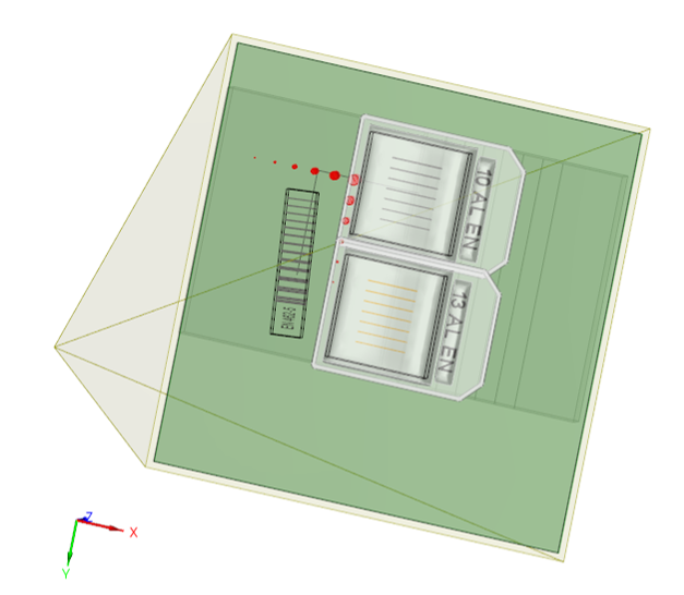
3D view of the model created in CIVA for the DURAL wedge with flat panel.
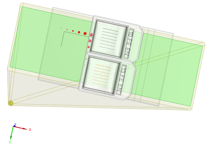
3D view of the model created in CIVA for the ferritic steel wedge with image plate.
Continue to Results
Back to Experimental protocol