
Computed Tomography with CIVA
The CT module proposes the same interface and same capatibilities as the RT Module in terms of:
- Specimens
- Sources (X-ray or Gamma-ray sources)
- Detectors
- Flaws
- Computation options: simulation of direct and scattered radiation
Simulations examples
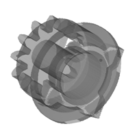
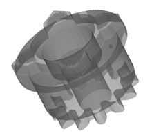
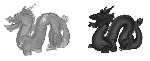
CT reconstruction obtained with CIVA
Specific items
The items specific to Computed Tomography are:
- Positioning
- Tomographic scanning
- 3D reconstruction
- Import of experimental data
Positioning
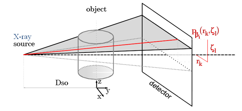
A positioning option allows the user to define the {detector-source} positioning system.
The user can now enter the source-axis distance and detector-axis distance in a semi-automatic way.
Specific misalignment options complete the positioning options. One can define an offset of the source and/or the detector to correct the misalignment versus the specimen to be inspected.
Tomographic scanning
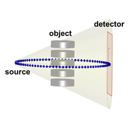
As the part geometry is fixed in CIVA, the X-ray tube and detector rotates around the specimen.
Different tomographic scanning can be modeled:
- A circular scanning for which the user defines an arbitrary number of steps and shot positions.
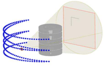
- A helical scanning, for which the user defines the length of displacement, the number of rotation, the number of positions per rotation and the maximal angle of rotation.
- A complex scanning (for instance robotic trajectories).
- A Linear scan of the source can also be performed, associated with linear detectors. It helps to reduce artifacts due to the source divergence, and to improve CT reconstructions with better quality for projections images.
Then, CIVA will run the RT simulation for all the projections.
Results
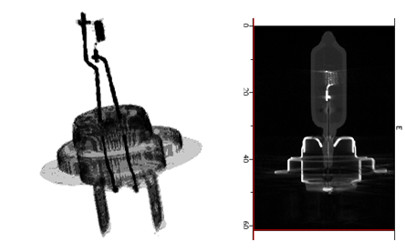
Results for each projection
X-ray slice data is generated for each position and the corresponding results can be displayed. Those results are identical to the RT module and depend on the detector and the computation options. All the analysis tools within CIVA RT are also available in CIVA Tomography.
CT data import
CIVA allows to import computed tomography experimental results from acquisition data. Different acquisition format are already available. If your format does not fit with CIVA import data tool, it is possible from some specific developments to ask to integrate this format thanks to plugins.
3D Reconstruction
Once the scan data has been acquired, the data must be processed using an algorithm of tomographic reconstruction, which produces a series of cross-sectional images.
The algorithms used in the current release of CIVA are the FDK algorithm (Feldkamp, Davis and Kress) for circular and helical tomographic displacement, PIXTV for circular tomographic displacement, and SART for complex trajectories.
- The FDK (Feldkamp, Davis et Kress) algorithm is a widely used filtered back-projection algorithm for three-dimensional image reconstruction.
- The PixTV algorithm is based on compressed sensing theory. This is an iterative reconstruction algorithm which minimizes the TV (total variation) norm.
Reconstructions using the FDK algorithm can be parallelized on CPU and GPU. For PixTV algorithm, reconstruction is realized on GPU.
Once the processing is finished, a new analysis page opens with the 3D reconstructed specimen. The 3D view available in CIVA shows the component in 3D dimensions. Many tools are then available to represent the reconstructed component: display of the iso surfaces, volume rending, etc.
On top of this, the user has the ability to see the different 2D sections and grab the different planes. All the sections and the 3D reconstructed view are linked together, by moving one cursor in one view, all the other analysis pages are refreshed in real time for a better interpretation of the results.