
Radiographic Testing with CIVA
The RT simulation module allows simulating a radiographic control by taking into account the different radiation produced by X or gamma ray sources.
From the simplicity of handling the CIVA interface, the user can easily and quickly set-up its configuration: Selection of the part to be inspected, definition and positioning of the source and the detector, insertion one or several flaws, definition of calculation options.
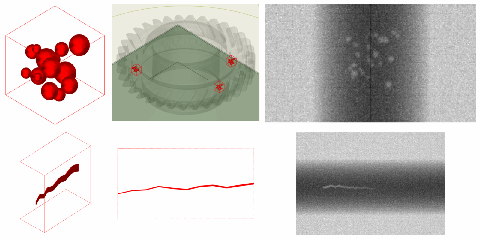
Simulation examples
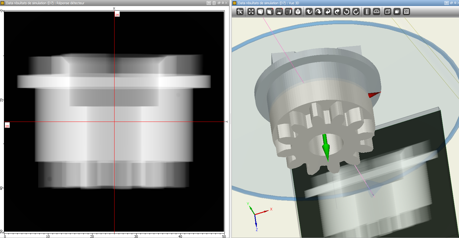
X-ray of a gear and display of the result in the 3D view
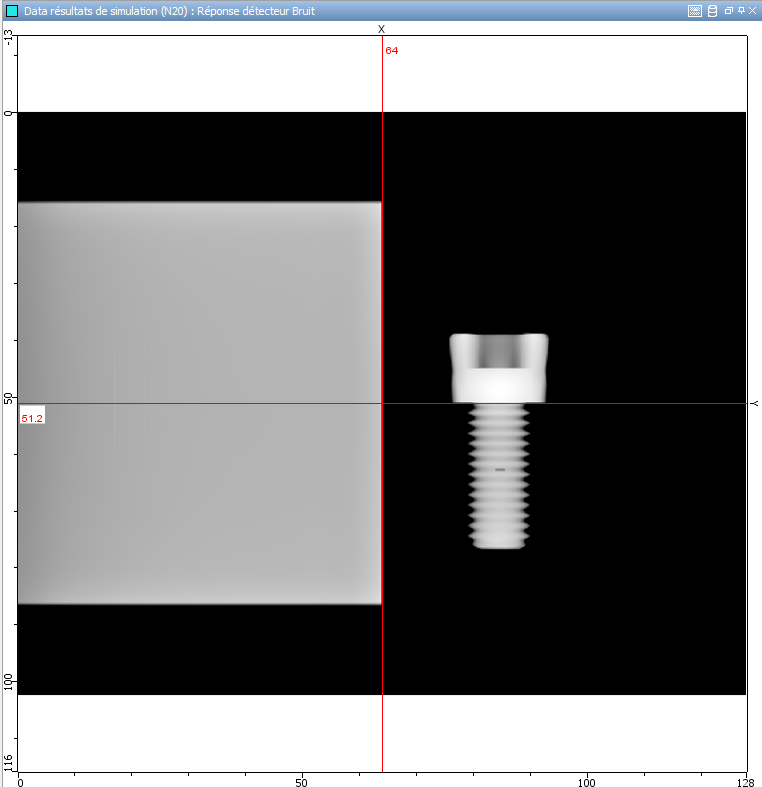
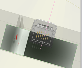
Radiography of a screw
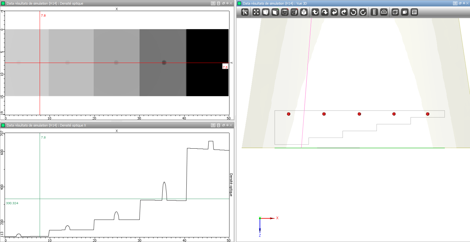
Radiography of a step wedge with display of the profile line along the X axis
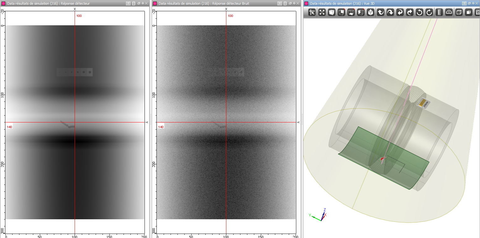
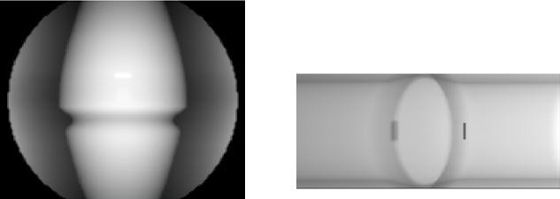
Radiography of a weld (display of results with and without noise)
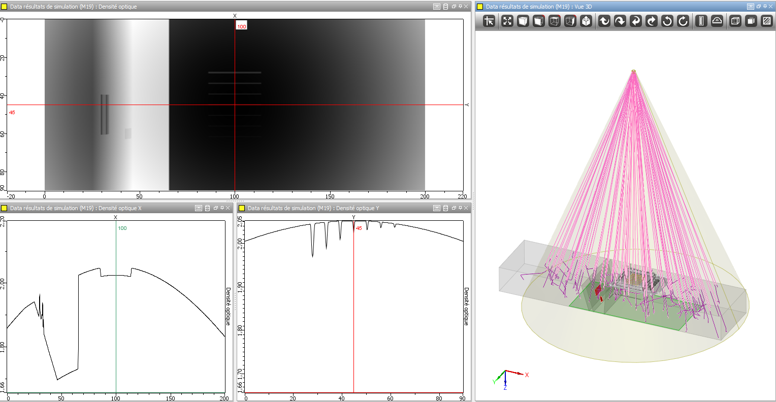
Radiography of a weld with display of the profile lines along the X and Y axis and display of the photon path in the specimen in the 3D view
Specimen
Parametric geometries and CAD files
The following specimens can be defined:
- Canonical specimens: flat, cylindrical, conical
- Preset components: Nozzle, butt or tee Weld (13 different bevel profiles available), turbine blade (blade root and groove), Through Wall Penetration, Elbow
2D profiles can be drawn or imported in CIVA (DXF format). The generation of the 3D geometry is effected by translation or by rotation of the profile. It may be homogeneous or heterogeneous.
3D CAD files (STL, IGES or STEP) can be imported in CIVA. Several volumes (through CAD and / or parametric geometries) can be imported simultaneously allowing to model complex geometries.
CIVA allows to export specimens modeled in IGES format.
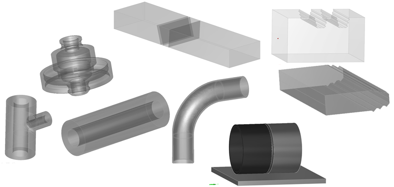
Examples of component geometries
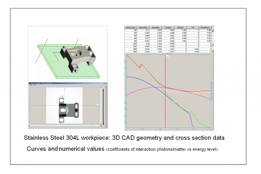
Materials
A library including over 110 elements and alloys is available in CIVA. The user can define, his own alloy by knowing the chemical composition of it. The new alloy can be named and saved for future use in CIVA. The component can be homogeneous (one material) or heterogeneous.
Source
X-rays sources, gamma-rays sources and high energies sources can be defined in CIVA RT:
- X-ray sources: Various parameters such as the intensity of operation, and the spectrum of the source must be defined. The spectrum can be defined in several ways:
- By selecting a predefined spectrum available in the library of source of CIVA
- By loading an experimental spectrum in CIVA (*.txt format file)
- By using a spectrum calculator included in CIVA based on the physical parameters of the source selected by the user: anode (angle and material) and acceleration voltage
- Gamma-ray sources: The activity of the source must be defined together with the radioisotope used. The classically sources used in NDT are already predefined: Cobalt 60, Iridium 192 and Selenium 75. Other sources can be added and saved in the library CIVA sources.
- High energies sources: A library of high energies spectra (linear accelerator and betatrons) is available in CIVA RT.
For X-ray and high energies sources, the user can add a filter to spectra.
Emission zone: It is possible to limit the effective zone of radiation in space with a conical or cylindrical volume. The effective size of the source (anode target with X-rays, radioactive capsule in Gamma) can be assumed as an ideal spot or not, allowing to account for geometrical unsharpness.
Detector
CIVA allows to model either digital and conventional radiography from libraries (based among others on the NF EN 11699-1 standard for silver films) or experimental data.
The detectors that can be modeled are:
- The silver films
- The image plates of high sensitivity or high resolution (with consideration of intensifying screens)
- Digital detectors type “flat panel”
- The CCD scintillators
A detector can be defined by simply keying-in its response in term of grayscale (or OD) in relation to a measured incident dose.
The detectors can be flat or curved, front and back filters can be added.
The detector noise or the granularity of the silver film can be taken into account. An MTF curve can be added for each type of detector.
A region of interest (ROI) can be used to over-sample the resolution of the computation at one part of the detector.
The radioactivity emitted by a specimen to be inspected can be taken into account by selecting the “Contact Dose Rate” option available for silver films. During the computation, this dose rate value will be added to the one emitted by the source modifying the result obtained.
A Photon counting detector (PCD) can be defined, making it possible to analyze and post-process the spectral distribution of incident photon energies thus providing interesting analysis features and image improvement capabilities.
Flaws
Several flaws can be inserted in the test piece. They can have different shapes: planar, spherical, ellipsoidal, trapezoidal, “crack-like”, or an arbitrary 3D CAD geometry. A cluster of porosities can be also defined in one shot. The same list of materials as for the specimen can be assigned for flaws.
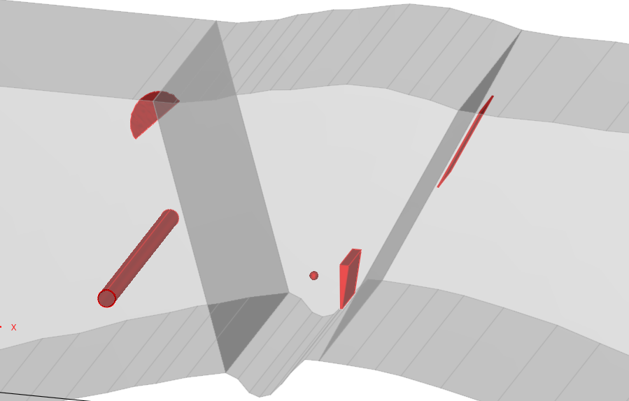
Examples of different types of flaws
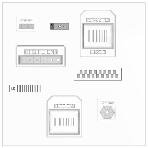
IQI
A library of the main standard penetrameters (based on EN, ASTM, AFNOR, DIN62, and CERL norms) is included in CIVA: wire (simple or duplex), holes, step wedge, etc.
Results
Two combined methods (analytical Beer-Lambert & Monte-Carlo) compute both the direct radiation and the scattered radiation. Scattering can include the following phenomena: Compton and Rayleigh interactions, Photo-electric absorption, pair creations, “Bremsstrahlung” effect (mainly relevant for high energy sources and thick specimen) and X-ray Fluorescence interactions in the specimen (mainly relevant for heavy materials). The build-up (ratio direct/scattered) is also available in order to estimate the importance of scattering in a given inspection. A scenario of parametric variations can be defined.
For Monte-Carlo computations, the algorithms benefit from multi-core architectures in order to reduce computation times. Moreover, if this computation has been performed once, and the user wishes to study the variation of a parameter that does not impact the scattering (variation of the exposure time for instance), the Monte-Carlo can be re-used in the new configuration and associated with a new and fast direct computation. CIVA includes a tool that will automatically provide the estimated number of photons for a good convergence of the Monte-Carlo simulation. It is also possible to continue a Monte-Carlo from a previous one if it is necessary to reach a better convergence for the scattering radiation.
A Linear Scan can be performed involving a scan of an X-Ray source used with linear detectors. This allows the user to reduce artifacts due to the source divergence and can be well adapted for long specimen inspections.
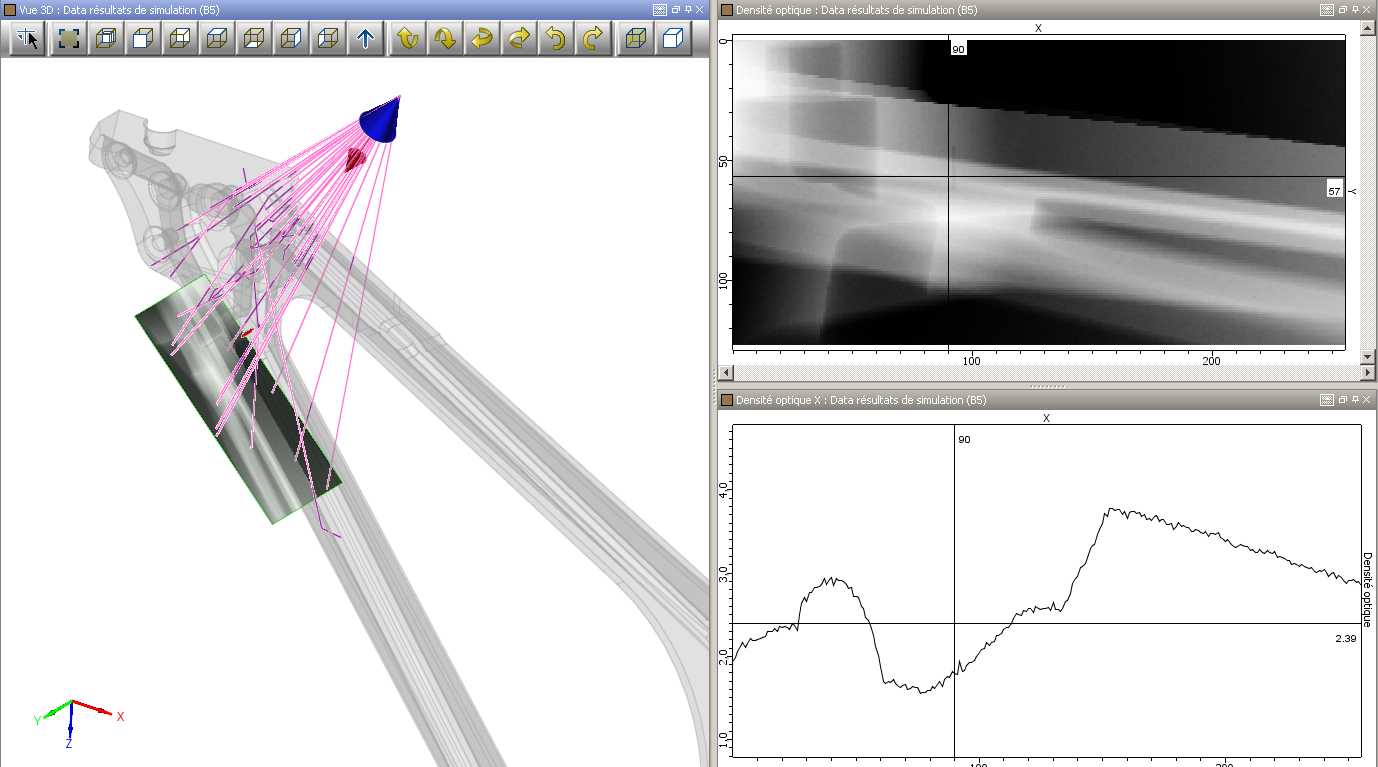
Result of a simulation of a stiffener radiographic testing: 3D view, photons path, image of the optical density, curves extraction
Result of two different pipes welding
The user can visualize both the detector response (optical density or grey levels) as well as the incident dose in Gy or the deposited energy on the detector in keV. The results are presented as images in the classical CIVA environment as well as curves following selected cross sections, allowing the user to easily quantify local variations of contrast. The cursors are dynamically linked to the 3D graphic view, and the photons paths are plotted. The thicknesses of materials passed through are identified in a table. The images obtained can be exported in TIFF or RAW format.
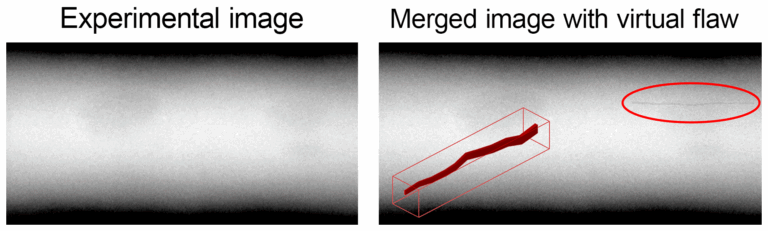
CIVA RT can also import experimental X-Ray images in TIFF format.
To provide even more realistic images, Data Fusion techniques have been implemented that let you insert virtual flaws (simulation results) into real images.
POD computations (Probability Of Detection) are also available in the RT module, based on the accounting of uncertain input parameters.
To understand and quantify the impact of influential parameters on an NDT inspection, parametric studies in CIVA are particularly adapted, since it is easy and fast to precisely change and monitor parameters. Based on a first set of computations, metamodels can also be calculated in CIVA, which provides extensive new results to the user in real time, as well as powerful analysis tools, such as multi parametric analysis and sensitivity analysis to evaluate the relative impact of influential parameters.
Post-processing capabilities are offered to the user, who can quickly re-calculate the activity of the gamma-ray source, the intensity of the X-ray source, or the relevant exposure time necessary to reach a targeted optical density (or response of detector). In the same way, the optical density (or response of detector) can be quickly recalculated if these 3 parameters change.
Some detectability criteria based on the smallest surface interpretable by the eyes have been implemented. These criteria will help the user to predict the flaw detectability. The detectability analysis is available for any simulations in CIVA RT.